Calcific Tendonitis
What is calcific tendonitis?

Calcific tendonitis of the shoulder happens when calcium deposits form on the tendons of your shoulder. The tissues around the deposit can become inflamed, causing a great deal of shoulder pain. This condition is fairly common although the cause is unknown and not related to injury, diet or osteoporosis. As well as the pressure caused by the calcium build up, the deposits reduce the space between the acromion and the rotator cuff leading to impingement (pinching of the tendons). Calcific tendonitis most commonly affects people over the age of 40.
There are two different types of calcific tendonitis of the shoulder: degenerative calcification and reactive calcification.
Degenerative Calcification
As part of the aging process, blood flow to the tendons of the rotator cuff decreases. This makes the tendon weaker. Due to the wear and tear as we use our shoulder, the fibres of the tendons begin to fray and tear, just like a worn-out rope. Calcium deposits form in the damaged tendons as a part of the healing process.
Reactive calcification
Why it occurs is not clear. It does not seem to be related to degeneration, though it is more likely to cause shoulder pain than degenerative calcification. Doctors think of reactive calcification in three stages.
- Pre-calcific stage: – the tendon changes in ways that make calcium deposits more likely to form
- Calcific stage: – the calcium crystals are deposited into the tendon. The deposits then begin to be absorbed by the body and disappear. This is usually when the pain will occur.
- Post-calcific stage: – the tendon heals and is remodeled with new tissue.
What are the symptoms of calcific tendonitis?
While the calcium is being deposited, you may feel only mild to moderate pain or possibly no pain at all. For some unknown reason, calcific tendonitis becomes very painful when the deposits are being reabsorbed, often constant and nagging. At times the pain travels down the arm to the hand and is aggravated by the lifting of your arm. The pain and stiffness of calcific tendonitis can cause you to lose motion in your shoulder. At its most severe, the pain may interfere with your sleep.
How will I know if I have calcific tendonitis?
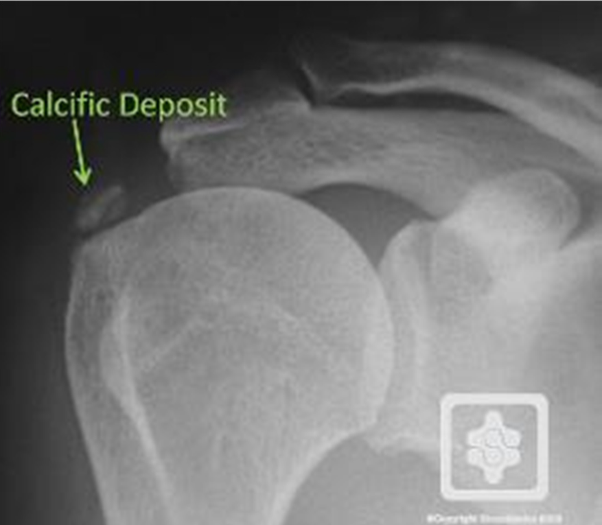
Your surgeon will take a detailed medical history and will conduct a thorough physical examination of the shoulder. An x-ray will usually confirm the presence of calcium deposits and will also help to pinpoint the exact location. Several x-rays may be required over time to assist the surgeon track the calcium changes and determine whether surgical is required.
Treatment Options
The main goal will be to reduce the pain and inflammation in the shoulder joint. Along with resting of the joint, the following options may be suggested:
- Anti-inflammatory Medications – These medications can be helpful in reducing swelling or inflammation in the shoulder joint, there in turn reducing pain.
- Injections – Cortisone is a very powerful steroid and can be very effective at easing inflammation and swelling. The effects are temporary, but can provide relief for several months.
- Lavage – During the time when the calcium deposits are being reabsorbed, the pain can be quite significant. Your surgeon may suggest trying to remove the calcium by inserting two large needles and rinsing the area with sterile saline solution. This procedure can loosen and break down the calcium particles which can then be removed with the needles. Even if the calcium cannot be removed, the lavage may reduce the pressure in the tendon, which can lessen the pain.
- Physiotherapy – Physiotherapy will also focus on reducing the pain and inflammation, which may include the use of ice or heat. The use of multiple ultrasound treatments to reduce the size of the calcium deposit is beneficial and may offer better arm function.
- Shock Wave Therapy – A machine is used to generate wave pulses directed at the painful area to help break up the calcium deposits so they may be more easily absorbed by the body. This treatment may also reduce the pain and the size of the deposits.
Do I need surgery?
If the loss of movement and pain continue to interfere with your daily activities your surgeon may recommend surgery. An arthroscopy (key hole surgery) is done where a small incision is made in the tendon and the calcium removed. Care is taken to ensure that the calcium has been completely removed by doing an x-ray at the end of the procedure. In very rare circumstances open surgery may be necessary.
What about Rehabilitation?
Rehabilitation after shoulder surgery can be a slow process. It is extremely important that you begin moving and exercising the arm following your procedure, unless otherwise specified. The physiotherapist will provide a program of strengthening and stretching exercises for you to do at home.
Even if you don’t need surgery, your surgeon may suggest that you attend physiotherapy sessions for 4 to 6 weeks. Strengthening the rotator cuff muscles can actually decrease the pressure on the calcium deposits in the tendon.
The patient information sheets are intended to provide general information only and are not a substitute for medical advice about your particular condition.